Sternal Injury
A less common chest wall injury, it is usually caused by a direct blow, associated with an acceleration-deceleration injury or from a fall onto the front of the chest. The Sternum or breastbone is a strong bone and in otherwise fit and healthily patients’ significant injuries to the sternum usually suggest a fairly severe injury or blow. However, in frail patients particularly with associated osteopenia (weakness of bones) even an apparent minor injury can cause a sternal fracture.
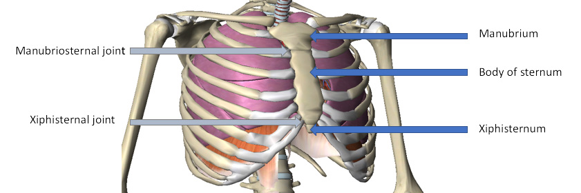
Anatomy of the sternum. The sternum is made up of three parts (blue arrows) with two junctions (grey arrows)
Symptoms
Patients with a sternal or breastbone injury typically experience a sudden onset of chest pain at the time of injury. Pain is often sharp and intense and may increase during deep breathing, coughing, laughing or sneezing. Patients may also experience an ache in the front of the chest that is particularly prominent at night or first thing in the morning (particularly in the first few days following injury). Pain may increase when lying down in certain positions (such as face down or on your side) and on firmly touching the sternum at the site of injury. Swelling and / or bruising may also be evident. In severe sternal fractures with bony displacement, an obvious deformity may be present. Patients with this condition may also experience pain with certain movements of the upper back and chest (such as twisting, bending forwards or sideways, or arching backwards) and with certain movements of the upper limb (such as pushing, pulling, heavy lifting or with overhead activities).
Diagnosis
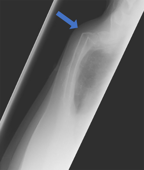
Sternal x-ray from the side showing significant sternal fracture.
As with rib injuries the diagnosis of a sternal injury is what doctors call a clinical one, that is taking a precise history of the injury coupled with a careful physical examination with a doctor familiar with chest wall injuries. Examination may demonstrate swelling, tenderness, occasionally crepitus (crunchy feeling under the skin) and if the sternum is badly broken a step. There is no specific blood test unless an associated chest infection or other internal complication is suspected. Radiological assessment with chest x-ray is not helpful as its very insensitive at picking up sternal injuries but may be helpful in identifying other associated problems such as fluid in the chest or a collapsed lung. More useful is a specific type of x-ray called a lateral sternal view.
If the injury is subtle occasionally a chest wall ultrasound may demonstrate a sternal fracture and associated bruising (haematoma) as well as identifying internal problems such as fluid around the heart (tamponade). The most sensitive radiological investigation is a Chest CT scan. This allows the severity of the sternal injury to be clearly seen as well as identifying any other chest related injuries such as haematoma behind the breastbone, fluid around the heart, lung bruising or contusions and other associated injuries.
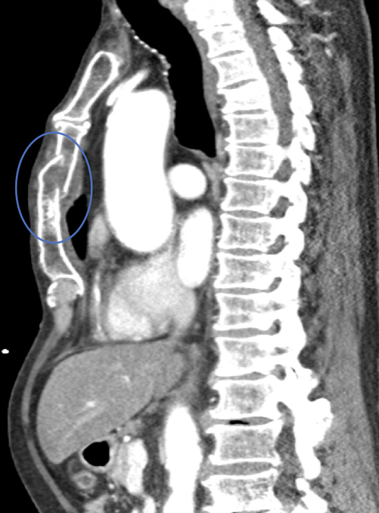
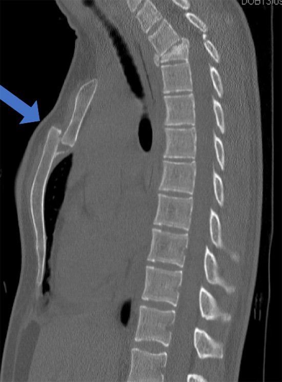
Video of a Chest CT scan taken from the side (sagittal) demonstrating a displaced fracture through the body of the sternum.
Severity
Sternal injuries vary significantly from a minor injury with associated pain, tenderness and bruising to severe sternal fracture. The type of injury and what happened is often the most useful guide to severity of sternal injury. One specific type of sternal injury is an acceleration-deceleration type of car accident in a seat-belted occupant either directly from a direct blow to the sternum or indirectly.
As with the classification of rib fractures, sternal fractures are based on the appearance on Chest CT scan. They are described typically as:
Simple: Usually a single fracture either partially or completely through the sternum, which is not displaced (dislodged) or angulated.
Chest CT showing an un-displaced sternal fracture albeit in two places (blue ring)
Complex: Usually single occasionally multiple and displaced where the broken ends are misaligned or overlapping with an associated step (lump) on the front of the chest.
Chest CT showing a displaced sternal fracture. The fracture is through the Manubriosternal junction and area of nature weakness of the sternum.
Complications
Pain: Immediate (acute) and can be severe over the area and is sore to touch and worse on certain movements. In most the pain will settle, however occasionally it can persist and become chronic causing significant issues. The main reason for this is failure to manage appropriately after the initial injury (with painkillers, rest, restrictions of activity and tailored return to normal activities).
Sternal fractures do generally heal even if displaced but the patient may be left with a permanent lump and tenderness.
Deformity: Occasionally if the sternum is badly displaced a tender lump can develop over the fracture site.
Breathlessness: Shortness of breath acutely is usually caused by the chest wall pain not allowing deep breaths to be taken, occasionally it can be associated with the lung collapsing after the injury; a build-up of fluid in the chest cavity (effusion) or even a developing chest infection (pneumonia). Chronically, on-going breathlessness can be due to chronic pain but also occasionally to complications of retained blood or fluid in the chest cavity which can trap the lung.
Internal injuries: Very rarely, if the sternal injury is severe internal injuries can lead to sinister symptoms of severe breathlessness and even collapse due to blood building up around the heart (tamponade) or bruising (contusions) of the heart itself. Diagnosis requires an ultrasound scan of the heart or chest CT scan.
Xiphersternal injury: Pain at the bottom of the sternum in the area of the 'solar plexus' can occur following even minor injuries and may be caused by an injury to the xiphersternal junction or the xiphersternum itself. The xiphersternum is actually made of cartilage and is susceptible to injury and inflammation leading to long-term pain and discomfort. For more information see Complex chest wall injuries.
Treatment
Sternal injuries can usually be treated conservatively involving rest, restrictions of activities and painkillers, or occasionally through some form of intervention including targeted physical therapy or surgical options such Open Reduction Internal Fixation. See Treatments.