Complex chest wall injuries
The Chest wall is a complex structure consisting of a bony skeleton with multiple joints between the sternum (breastbone) and vertebrae (spine), and between the bony parts of the ribs and the costal cartilage part of the rib cage at the front of the chest between the ribs and sternum. In addition, there are multiple ligamentous, tendinous and muscle attachments. Many of this ‘junctional’ areas can be sources of injury or prone to inflammation and damage.
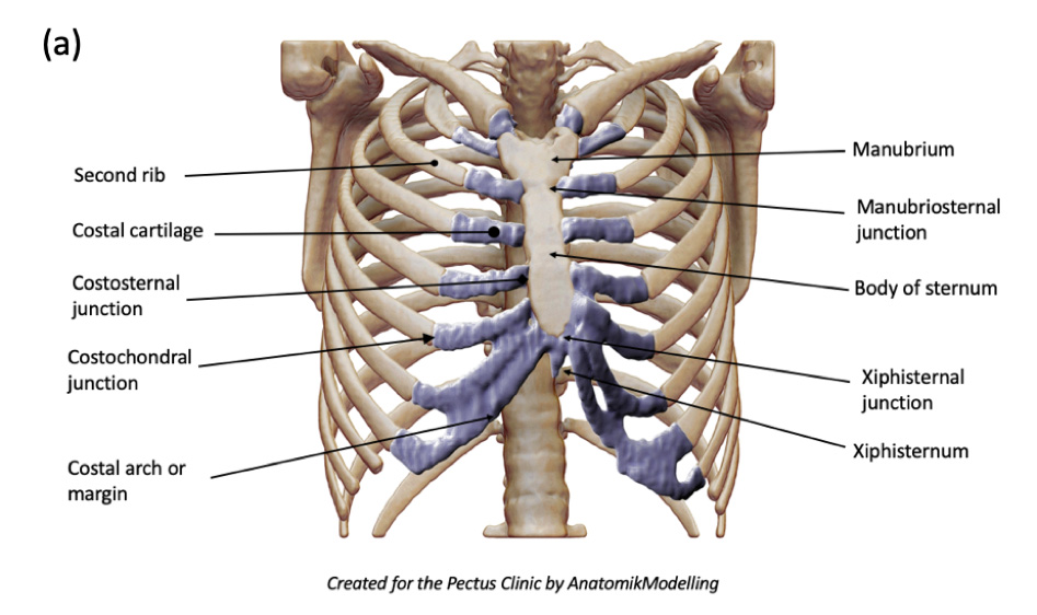
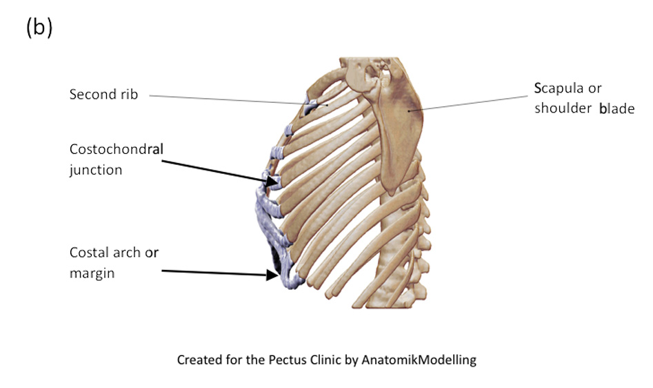
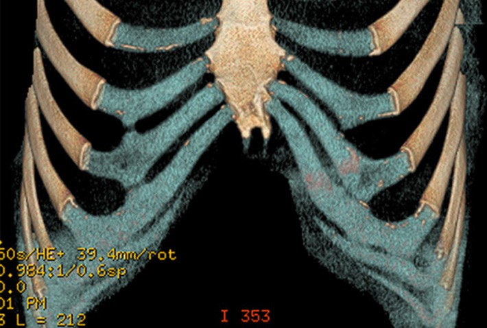
Annotated image of a CT 3D reconstruction showing the (a) anterior and (b) lateral chest wall structures.
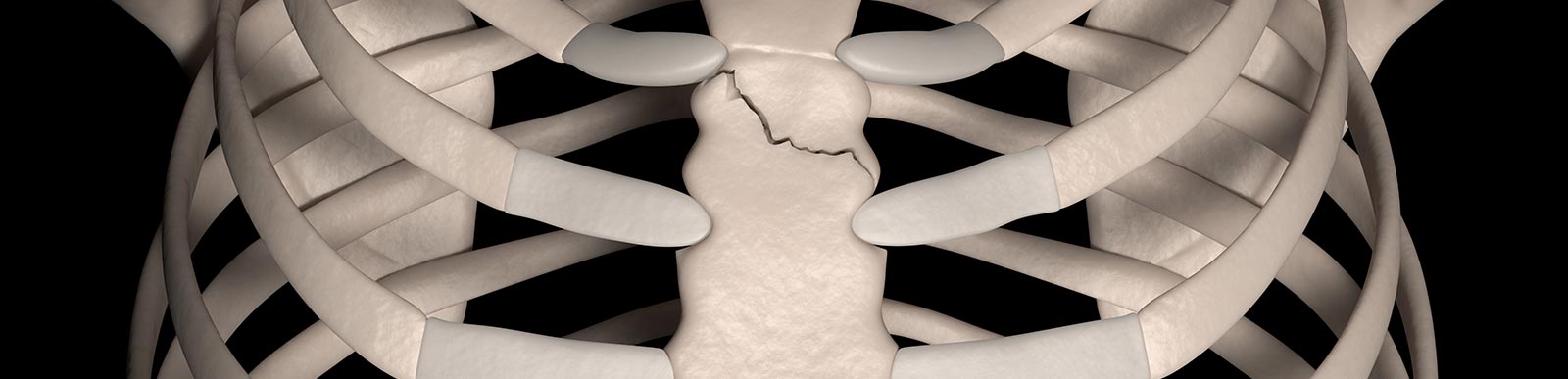
Manubriosternal junction
Refers to where the manubrium sterni (top of sternum) and body of sternum joint together with the 2nd rib. It is a fibrocartilaginous joint and easily palpable as an often raised prominence as the manubrium joins the sternum at a slight angle (referred to as the sternal angle).
Symptoms
The maubriosternal junction can be injured through a direct blow or indirectly through acceleration-deceleration compressive-decompressive type of injury (for example during a front on collision in a seat belted passenger). This results in a dislocation and can lead to severe pain, tenderness and swelling (a step) in the area. If such an injury is suspected a chest CT scan is required to diagnosis and to assess for other chest injuries.
A more insidious problem is the development of localised inflammation in the area leading increasing pain, discomfort and occasionally the area can be hot or swollen. A high clinical suspicion is regarded and together with radiological investigations such as an MRI.
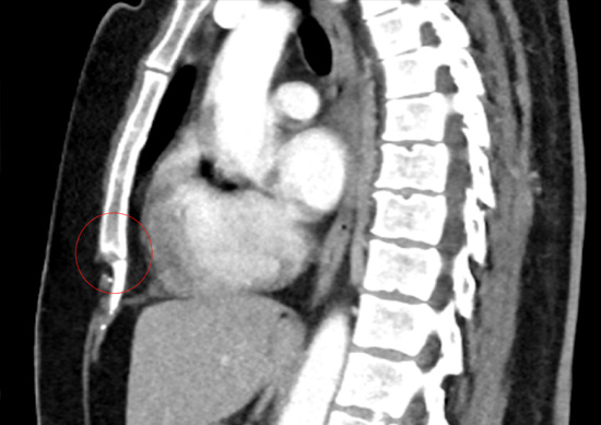
Manubriosternal junction or Sternal angle (highlighted with red circle)
Costosternal junction
Ribs can be classified on the basis of their costosternal attachments: true ribs if their costal cartilage attaches directly to the sternum, false ribs if their costal cartilage attaches to the cartilage of the next most superior rib (as part of the costal arch), and floating ribs if there is no costal cartilage connection. Ribs 1–7 are true ribs, ribs 8–10 are false ribs, and ribs 11 and 12 are floating ribs.
Costochrondral junction
Refers to the junction or joint between the ribs and the costal cartilage at the front of the chest typically away or lateral to sternum. Like the costosternal joint, the upper 8 costochondral junctions forms a type of joint (hyaline cartilaginous joints) which are more like the junction between the first rib and sternum (synchondrosis) so form a tough type of joint very little movement. The 9th and 10th rib costochondral junctions are fibrous.
Symptoms
The main types of pain seen are due to either injury from a blow or indirect trauma and inflammation.
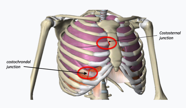
Illustration of Costosternal and costochondral junctions
Acute trauma to costosternal / costochondral joint from an injury to the front of the chest such as a direct blow, or indirectly. It can be quite minor such as a twisting injury whilst lifting or even a violet cough or sneeze. It is typically associated with sudden and severe pain in the area next to the breastbone at the level of the injury or further away from the sternum with costal cartilage meets its rib connection. The pain is likely to be tender over the spot, and can be associated with swelling, even bruising and occasionally a sense of movement or even ‘popping’ caused by subluxation (partial disconnection of the junction) or dislocation (complete disconnection).
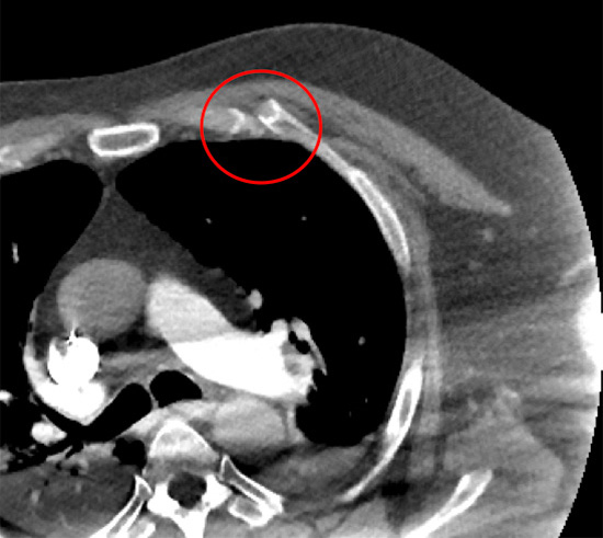
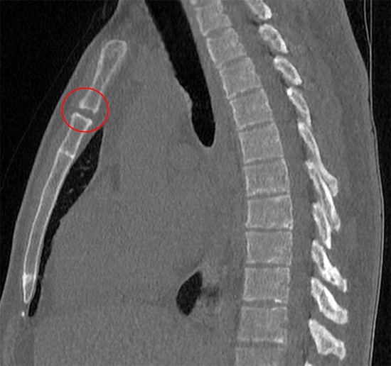
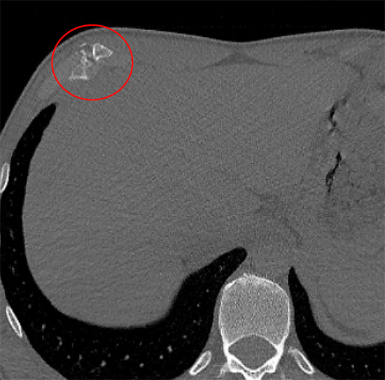
Chest CT scan following fall of an acute costochondral facture (red circle)
Costochondritis (Inflammation of the cartilage junctions of the sternum or ribs) and Tietze syndrome (sudden chest pain and localised swelling at junction of the sternum and ribs) are inflammatory causes of sternal and chest wall pain, see Costochondritis and other inflammatory problems.
Costal arch
Costal arch or margin refers to the lower edge of the chest formed by the bottom edge of the rib cage. It is formed by the 7th to the 10th rib costal cartilages to create the costal arch. Interchrondal junctions or articulations are joints between costal cartilage forming the costal arch and the lower ribs. The costal arch because it is made of cartilage is actually quite difficult to image.
CT reconstruction and rendering of the costal cartilage (in blue) showing the costal arch or margin
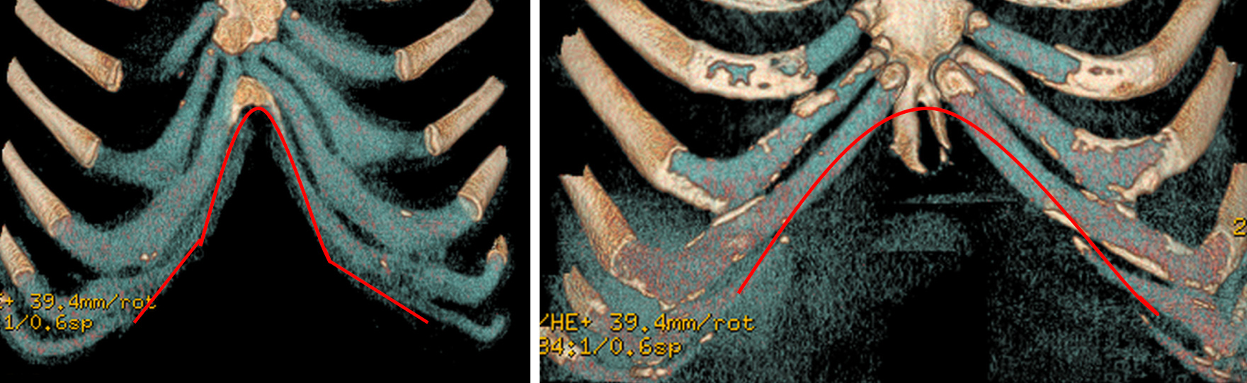
The costal arch varies in shape with narrow (left) and broad wide costal arches (right). Age, gender, obesity (particularly truncal abdominal obesity (pot belly) influence the shape
There appears to be some variation between how the costal cartilage of the lower ribs particularly the 9th and 10th ribs articulate with the costal arch. If the lower costal cartilages are not fully attached to the costal arch it may lead to a condition called slipped rib syndrome.
The shape of the lower chest created by the costal arches is quite variable with variation seen. Rib flare refers to a particular type of costal arch that is prominent particularly on the left and is often but not always associated with pectus chest wall deformities.
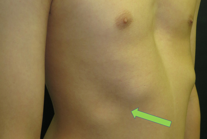
Bilateral rib flare showing a right sided costal arch with a dip (or deficit) (arrow in green) where the lower costal arch has not formed completely. In this case it was associated with slipped rib syndrome. NOTE: Incidental pectus Excavatum.
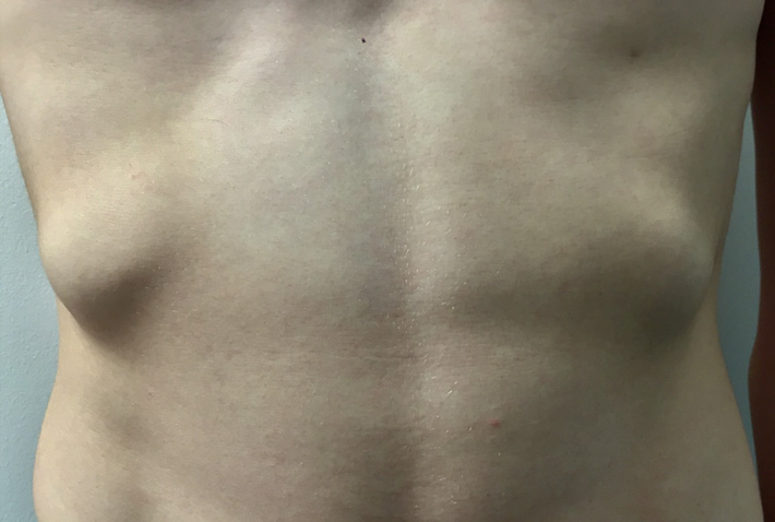
Bilateral rib flare in a young man with no symptoms or other issues
Symptoms
The main types of pain seen are due to either injury from a blow or indirect trauma and inflammation.
Acute trauma to costal arch from an injury to the front of the chest such as a direct blow, or indirectly. As with costosternal and costochondral injuries it can be quite minor such as a twisting injury whilst lifting or even a violet cough or sneeze. It is typically associated with sudden and severe pain in the area of the costal arch. The pain is likely to be tender over the spot, and can be associated with swelling, even bruising and occasionally a sense of movement or even ‘popping’ or even a ‘step’ caused by subluxation (partial disconnection of the junction) or dislocation (complete disconnection).
Old fracture of the costal arch leaving a ‘lump’ and chronic pain (red circle)
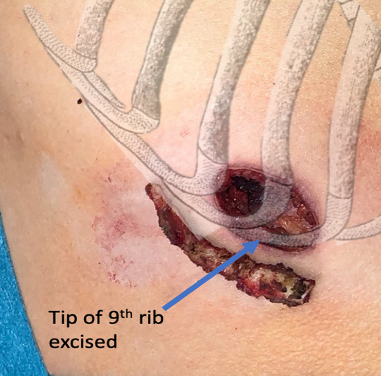
Left costal arch with excision of 9th rib tip (with costal arch illustrated in grey)
Slipped rib syndrome refers to pain of the lower costal arch caused by abnormal movement of the lower costal cartilage (typically the 8th, 9th or 10th rib tips). It’s likely to be associated an abnormal or ‘lose’ connection between articulations of the costal arch which then allows movement leading to irritation and pain. See Slipped Rib Syndrome.
Xiphersternal junction
At the bottom of the breastbone (sternum) is a further junction between the bony bottom of the sternum and the cartilaginous xiphisternum or xiphoid process. Xiphersternal junction is a type of joint with very little or no movement.
Symptoms
Pain caused by the xiphoid process is called xiphoidalgia. Xiphoid process pain occurs for varying reasons but is often related to minor trauma to the area, over exercise (such as abdominal crunches) or can become more prominent following excessive weight loss. Local inflammation can also occur. See Costochondritis and other inflammatory problems.
Chronic xiphersternal junction fracture (red circle) following direct blow leading to a lump and chronic pain on exertion
Treatment
Complex chest wall injuries can be treated either conservatively involving rest, restrictions of activities and painkillers, or through some form of intervention including targeted physical therapy, trigger point injections or a variety of surgical options. See Treatments.